Why Electron Microscopy Crucial in Renal Biopsy.
Why Electron Microscopy is Crucial in Renal Biopsy?
When doctors suspect something is wrong with your kidneys, one of the most valuable tools they use to find out exactly what’s going on is a Renal biopsy. In this procedure, a tiny piece of your kidney is examined under different types of microscopes to identify the cause of the problem. One of the most powerful microscopes used in this process is the Electron microscope.
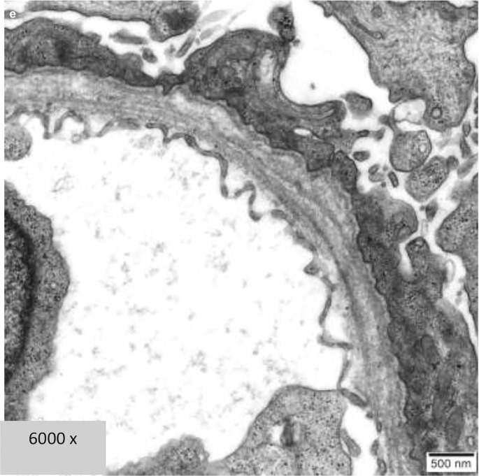
Electron microscopy is a special technique that allows doctors to look at the kidney in ultra-fine detail. While regular microscopes can only show cells up to a certain level of magnification, electron microscopes zoom in much further, showing the structure of the kidney at a molecular level.
So, why is this level of detail important?
The kidneys are made up of tiny filtering units called Nephrons, and each nephron has even tinier parts, such as capillaries, tubules, and the glomerular basement membrane (which helps filter blood). Many kidney diseases affect these structures in ways that are invisible to standard microscopes, but an electron microscope can reveal those subtle changes.
This additional detail is sometimes crucial for reaching the right diagnosis, especially when the cause of kidney damage isn’t obvious. It’s like trying to understand what’s wrong with a clock: if you look at it from a distance, you might see it’s not working, but you wouldn’t know whether the problem is with the gears, the hands, or the springs. Electron microscopy helps doctors look “inside the clock” to determine the exact problem.
When Might Electron Microscopy Be Important in a Renal Biopsy?
In several situations, electron microscopy becomes a game-changer in diagnosing kidney diseases. Many kidney conditions, such as Glomerulonephritis, Minimal change disease, and Alport syndrome, and other diseases affect the microscopic structures of the kidney subtly. Without electron microscopy, these conditions may go unnoticed or be misdiagnosed.
Here’s a real-world example that highlights its importance :
A Case of which changed the course of disease – by doing the electron microscopy.
A 35-year-old patient presented with swelling in the legs and feet, along with significant protein in the urine (proteinuria). Initially, the kidney biopsy was examined under a regular light microscope, and the findings suggested Focal segmental glomerulosclerosis (FSGS), a disease where parts of the kidney’s filtering units scar over time. The patient was started on steroids, a common treatment for FSGS, but there was no improvement after several months.
At this point, the medical team decided to re-evaluate the biopsy using electron microscopy. The results were eye-opening. The ultra-detailed view showed that the kidney’s glomerular basement membrane was not scarred, as seen in FSGS, but rather showed changes more consistent with Fibrillary Glomerulonephritis” it is is a condition characterized by the production of abnormal proteins that clog the glomeruli and cause inflammation.
The diagnosis was changed from FSGS to Fibrillary Glomerulonephritis, and the patient’s treatment was adjusted accordingly. The prognosis for FGN is poor, with nearly half of patients progressing to end-stage kidney disease within 2 to 4 years. Even with a renal transplant, there’s a high recurrence rate.
Key Takeaway :
The example above highlights just how important electron microscopy can be. While light microscopes are essential for diagnosing many kidney conditions, they have their limits. Electron microscopy allows doctors to see details that might otherwise be missed, leading to more accurate diagnoses and more effective treatments.
For patients with kidney disease, this can make a big difference. An early and correct diagnosis means the right treatment can begin sooner, which can prevent further damage to the kidneys and improve long-term outcomes.
Read More About : https://www.drsuhasmondhe.com
www.kidney.org/kidney-topics/kidney-biopsy
Conclusion :
While the idea of using an electron microscope might sound like something out of a science fiction movie, in reality, it’s a vital part of kidney care today. This high-tech tool allows nephrologists to see kidney disease on a molecular level, helping them make more accurate diagnoses and improve patient care. If your doctor recommends electron microscopy as part of a kidney biopsy, rest assured it’s because they want to give you the best possible understanding of your condition.
By using every tool available—especially electron microscopy—kidney specialists can ensure you get the right diagnosis and the most appropriate treatment, improving your chances of a healthier life with well-functioning kidneys.


Leave a Reply